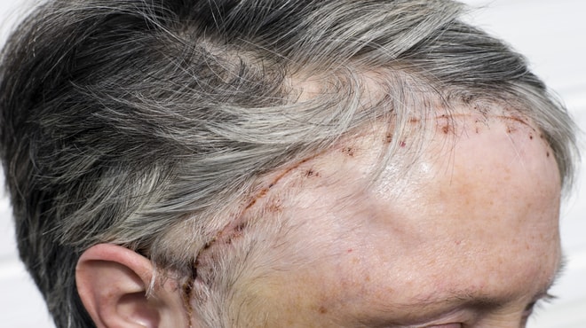
Surgery refers to the removal of tumor tissues in the brain and in the surrounding regions to which it has spread locally. Surgical procedures for brain depend upon the type and grade of the tumor. The following are some brain cancer surgeries doctors perform:
Craniotomy:
The surgeon first remove a piece of bone from the skull for access to the tumor, using specialized tools. Then, we remove all or most of the tumor. After surgery, bone replacement takes place. In a craniotomy, replacement occurs later (or not at all) for various reasons, like if swelling persists after surgery.
Endonasal Endoscopy:
We use an endoscope to navigate and access tumors through the nose and sinuses. Using image guidance and special instruments attached to the endoscope, this type of brain cancer surgery allows us to then remove tumors or take samples for biopsy.
Neuroendoscopy
Through a small opening in the skull, we use an endoscope to see inside the brain. We can then remove tumors or take biopsy samples using special instruments attached to the endoscope.
Other Procedures
Shunt: We place a thin tube (called a shunt) into a ventricle of the brain, through a small hole in the skull. The shunt moves excess fluid from the brain to another part of the body. For instance, in the abdominal cavity, where it is absorbed into the bloodstream. A filter catches stray tumor cells that may be in the cerebral spinal fluid (CSF). This procedure can help relieve pressure in the skull.
Placement of an Ommaya Reservoir: During this brain cancer surgery, we implant a small reservoir attached to a tube under the scalp. The tubing leads into a ventricle of the brain where the CSF circulates. This allows for chemotherapy administration to the brain and CSF, or to remove fluid for biopsy. The reservoir can be removed when it’s no longer needed.
Surgical procedures for brain cancer
There have been rapid advances in surgery for brain tumors, including the use of cortical mapping, enhanced imaging, and fluorescent dyes. Depending on the type and grade of the tumor, the following are some brain cancer surgeries our doctors perform.
- Cortical mapping helps identify areas of the brain that control the senses, language, and motor skills.
- Enhanced imaging devices give surgeons more tools to plan and perform surgery. For example, computer-based techniques help surgeons map out the location of the tumor very accurately. However, this is a very specialized technique that may not be widely available.
- A fluorescent dye (5 aminolevulinic acid) is orally given before surgery. This dye taken up by tumor cells helping doctors safely remove as much of the tumor as possible.


