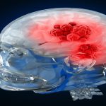
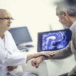
When the patient presents with suggestive symptoms of brain tumor, the physician will do a thorough medical history and physical examination. Then a plan for screening tests and procedure is drawn out for diagnosis of brain tumor
Your doctor may decide to do some of the following tests:
- Physical exam
- Mental assessment
- Eye exam or eye test
- Hearing tests
- Testing your facial muscles
- Testing your swallowing or gag reflex
- Checking the strength in your limbs
- Checking your balance or coordination
- Checking the sensation on your skin
Imaging can be done by the following ways:
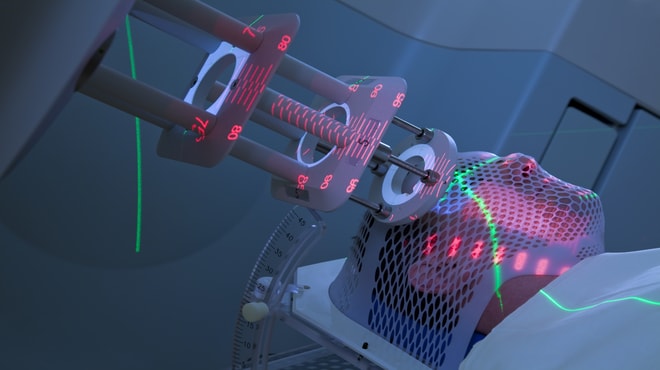
1. Computed tomography (CT) scan.
A CT scan creates a three-dimensional picture of inside the body using X-rays that are taken from different angles. A special kind of dye is also used to make the image clearer. The dye can be injected into the patient’s veins or can be given as a liquid to swallow. However, even in a CT scan, tumors that are very small are difficult to recognize.
CT angiography (CTA):
Here, the patient is injected with a contrast dye through an IV line while the scanner is at work. This creates detailed images of the blood vessels in the brain helping doctors to plan surgery.
2. Magnetic Resonance Imaging (MRI).
The MRI scan unlike the X-ray uses magnetic waves to give a detailed image of the internal parts of the patient’s body. The MRI is also known to detect the tumor size and a dye called a contrast medium is injected into the patient’s veins to give a more detailed view.
Special types of MRI are as follows:
a. Magnetic resonance angiography (MRA) and venography (MRV)
Used to get images of the blood vessels, this is done during or before surgery.
b. Magnetic resonance spectroscopy
Done as a part of MRI, MRS measures biochemical changes in any area of the brain.
c. Magnetic resonance perfusion
A special type of MR image is taken to determine the amount of blood going to different parts of the brain after a contrast dye injection. This indicates the site of the tumor as a faster growing tumor may need more blood. This in return helps determine which tissues need biopsy to be done.
d. Functional MRI (fMRI)
This test determines the functions different regions of the brain handle so that doctors avoid these regions while planning treatment modalities.
3. Biopsy
A biopsy is a procedure where a sample tissue is removed and looked at under a microscope to see if it is cancerous. Biopsies can be done through different methods like needle biopsy, laparoscopic biopsy and surgical biopsy. For brain tumors, the biopsies are of two types:
a. Stereotactic biopsy
This type of biopsy is done based on imaging tests.
b. Surgical or open biopsy (craniotomy)
If imaging tests confirm that the tumor exists, the neurosurgeon may not do a needle biopsy. Instead he may remove tumorous regions through a surgery called craniotomy. Removing most of the tumor is called debulking.
4. Positron emission tomography (PET) scan
The patient is injected with a mildly radioactive substance (usually a sugar called FDG) and this collects mainly in tumor cells. A camera that is of less quality than CT and MRI scans helps build information for further action.
5. Positron emission tomography (PET) scan
A chest X-ray looks for tumors in the lung if a tumor is found in the brain. This is because many instances of metastasis into the brain have been reported of lung cancers.
6. Electroencephalography
An EEG is used for a measurement of electrical activity of the brain. It also makes it possible to monitor seizures.
7. Evoked potentials.
Like EEG’s, involve electrodes that measure electrical activity; only those of the nerves. They often detect acoustic schwannoma, a noncancerous tumor.
8. Lumbar puncture or spinal tap.
In this procedure, the doctor uses a needle to take sample of cerebrospinal fluid (CSF) to look for tumor cells, blood, or tumor markers.
9. Myelogram
This is used to find if the tumor has spread to the spinal fluid and other parts of the brain.