
Here’s a look at the most common types of breast cancer imaging screening tests available today. Apart from these, breast cancer self-examination (self checking for lumps) and clinical breast examination (breast examination by treating physicians) are also important in catching cancer in the early stages, while it is still easily treatable.
The goal of breast cancer screening is to find cancer early, before it has a chance to grow, spread, or cause problems.
Early detection of breast cancer through screening procedures reduces the risk of death from breast cancer, and increases your treatment options. In the early stages, treatment options include less extensive surgery and/or the use of chemotherapy with fewer side effects, and sometimes you might even have the option of avoiding chemotherapy. Such options do not exist for the advanced stages.

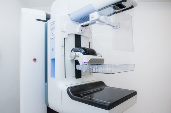
A mammogram is an x-ray picture of the breast that aids in the early detection and diagnosis of breast diseases in women. Mammography is the most common screening test for breast cancer. Other screening methods include Breast MRI, ultrasound breast and clinical breast examination.
Types of mammography
Conventional Mammography
Also known as film mammography, this type uses X-ray radiation to produce an image of the breast. Conventional mammograms are read and stored on film, which may result in false positives or an inaccurate diagnosis.
Digital Mammography
This is also known as full-field digital mammography or FFDM. A digital mammography machine takes digital X-ray images of breast tissue that can be magnified or enhanced to make the evaluation of the x-ray a lot easier, thus producing more accurate results.
Digital mammogram images are stored on a computer and can be transmitted electronically.
Digital mammography uses less radiation and also has better picture clarity. This helps in detecting abnormal changes more accurately. This creates fewer false positive test results.
3D Mammography
3-D mammography is a state-of-the-art 3-D breast imaging technology, also called breast tomosynthesis or digital breast tomosynthesis (DBT).
It is an advanced form of breast imaging that uses x-rays to take a series of pictures of the breast from many different angles.
A computer is used to make 3-D pictures of the breast from these x-rays.
This was FDA approved in 2011 for breast cancer screening.
Breast tomosynthesis has the following advantages over conventional mammography
- While DBT has been shown to have benefits for all patients, it is especially beneficial for individuals who are at high risk for breast cancer, either by genetic predisposition or by family/personal risk factors.
- It helps in the early detection of small breast cancers that may remain hidden on a conventional mammogram.
- Clearer images of abnormalities within dense breast tissue are possible with this technology.
- Greater accuracy in pinpointing the size, shape and location of breast abnormalities becomes possible.
- Fewer unnecessary biopsies or additional tests will be required after a 3D mammography has been done.
- There is increased likelihood of detecting multiple breast tumors
Though it is better than standard 2D mammograms in terms of safety and accuracy, the drawbacks are that they cost more than traditional mammograms and are available at very limited centres.
Why do we need mammograms?
Usually, mammograms are required for two purposes: screening and diagnosis.
Screening Mammograms
The screening mammogram is a routine breast examination process. It is used to detect signs of breast cancer in women who have no symptoms.
- These mammograms make it possible to detect tumors that cannot be felt.
- The machine takes pictures of the breast from two angles. It usually takes only 10-15 minutes for the scanning of the breast.
Sometimes even a routine screening mammogram can reveal abnormal growth in the breast tissue. While the growth can be benign (meaning harmless), it could also raise suspicion of being cancerous.
Diagnostic Mammograms
The diagnostic mammogram may be recommended by your oncologist if he/she detects any abnormal growth in the breast that could be a sign of cancer.
- This process works a little differently from the screening mammogram and could take longer too.
- These are used to diagnose abnormalities found in a screening mammogram or when other symptoms have been found (lumps, nipple pain, discharge).
- These take longer to perform than a screening mammogram.
- The dosage of radiation is higher because more x-ray images of the breast tissues may be required, especially where the abnormal growth has been spotted. This is done in order to precisely determine any change in shape or thickness as well as to take a closer and deeper look at the suspected tumour.
- Additional imaging may be necessary, such as an ultrasound or biopsy
When should you get a mammogram?
Recommended screening guidelines are dependent on your health history and your risk for developing breast cancer. Speak with your doctor to determine when you should start getting screening mammograms.
Women with average risk of breast cancer should start getting mammograms between ages 40 to 45. Regular screenings, which typically occur once or twice a year, should continue as long as your doctor recommends.
The schedule for screening might also be different if you have a high risk of breast cancer. You can talk with your doctor about how often you should have a mammogram based on your risk as well as your preferences. For example, you might opt for regular mammograms even if you are under the age of 40, because you might have a relative who was diagnosed with breast cancer at a young age, or if you have certain genes that increase your risk of breast cancer (such as “BRCA” genes).
In addition, your doctor may recommend a mammogram for the following reasons:
- To diagnosis breast irregularities you or your doctor finds during a breast exam
- To follow-up on a previous mammogram that showed some abnormal results
- To track the progress of lumps or other irregularities.
What to expect during a mammogram
When you go to the medical centre for the scan, you will be required to wear a loose gown. Most scan centres provide this type of garment.
The technician will assist you in comfortably positioning your body and breasts in the right way. Each breast will be individually scanned.
While a tray supports the breast, a flexible plastic plate is gently pressed down on top of the breast and against the breast tissues. You may feel slight discomfort but that is only natural.
The machine’s tube moves in an arc over the breast from various angles as it takes several low-dose X-rays of the breast tissues in a continuous pattern, and transmits extremely clear images to a computer.
The computer puts these digital images into a series or layers of thin slices. These are then stitched together to give high resolution images of the breast and the tissues.
The entire process involving both breasts takes only a few seconds each.
Getting a mammogram can be uncomfortable, but it lasts only a few seconds. If your breasts are sensitive before or during your period, you might want to avoid scheduling your mammogram during that time, if possible.
Do not use underarm deodorant or powder on the day of your appointment.
Do not delay
If you have been experiencing something abnormal in your breast, do not ignore the feeling.
Not all pains or lumps in the breast are indicative of cancer. Even if there is a lump, it may not always be cancerous. But there is no way to know, except to visit a doctor and get a proper diagnosis.
The more you delay, the more the cancer grows and moves into advanced stages. When cancer spreads to other organs, it becomes more challenging to treat it successfully.
Visit your doctor at the earliest and speak about getting a mammography done.
Still confused? Call our care manager on 79965 79965 to find answers.
You can read more about other types of cancer screening here.