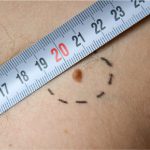
Skin cancer can develop in any part of the body, but most often it is developed on skin that is more exposed to the UV radiation. Skin cancer occurs to people of all skin tones. Read more about skin cancer here.
How is skin cancer staged?
Staging standardizes the process of describing how much the cancer has spread in the body. Staging of cancer helps the doctors figure out how much the cancer has spread and determine its best treatment and also helps calculate survival statistics. The lower the number of the stage, the less is the cancer has spread, with early stages being 1 and the most advanced stage being 4.
TNM method:
Most cancers that have tumours are staged using a staging system called TNM system and the same is used for skin cancers. The size of the primary tumour (T), the presence of cancerous lymph nodes (N) and how far the skin cancer has spread to a different part of the body (M) can be described using the TNM system.
High risk features:
The following are the high risk factors of skin cancer:
- The cancer is more than 2 mm thick in diameter
- It has grown into the lower dermis
- It has grown into the space around a nerve
- It started to spread to the ear or lip
- The cells are poorly differentiated or undifferentiated when seen in a microscope
Read more about risk factors of skin cancer here.
Stage I non melanomas:
Basal cell carcinoma is rarely staged as these are almost always cured before they spread to other parts of the body, however in the cases where it needs to be staged, TNM method of staging is used. Squamous cell carcinomas are staged similarly.
The cancer is 2 cm across or lesser and has one or no high risk features.
Treatment of stage I basal cell carcinomas:
The following are the treatment methods available for treating localized basal cell carcinomas:
- Simple excision
- Mohs micrographic surgery
- Radiation therapy
- Curettage and electrodesiccation
- Cryosurgery
- Photodynamic therapy
- Topical chemotherapy
- Topical immunotherapy
- Laser surgery
Treatment of stage I squamous cell carcinomas:
The following are the options available for treating localized non melanomas.
- Simple excision
- Mohs micrographic surgery
- Radiation therapy
- Curettage and electrodesiccation
- Cryosurgery
- Photodynamic therapy
Stage I melanoma:
Stage I of melanoma is further divided into two stages IA and IB.
Stage IA:
Stage IA is characterized by the cancer being less than 1 mm thick, the skin covering the cancer is not broken (not ulcerated) and the cancer has a mitotic rate of less than 1/mm^2.
Stage IB:
Stage IB is characterized by the cancer being less than 1 mm thick but the skin is ulcerated or the thickness being between 1 mm and 2 mm but the skin is not ulcerated. The cancer has mitotic rate being more than 1/mm^2.
Treatment of stage I melanoma:
Surgery is the main treatment option for stage I melanoma, the tumour and a small areas of surrounding skin being removed may be sufficient for removing the cancer completely at this point. A second operation to remove a larger area around where the melanoma was and this is called wide local excision. A sentinel lymph node biopsy may be done to check if the nearby lymph nodes are affected.


