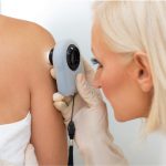
Skin cancer can develop in any part of the body, but most often it is developed on skin that is more exposed to the UV radiation. Skin cancer occurs to people of all skin tones. Read more about skin cancer here.
How is skin cancer staged?
Staging standardizes the process of describing how much the cancer has spread in the body. Staging of cancer helps the doctors figure out how much the cancer has spread and determine its best treatment and also helps calculate survival statistics. The lower the number of the stage, the less is the cancer has spread, with early stages being 1 and the most advanced stage being 4. The staging of skin cancer depends on the type of skin cancer and even the location of it too.
Types of skin cancer:
Skin cancers can be divided into two main categories, non melanomas and melanoma. The types of skin cancers are:
- Basal cell carcinoma
- Squamous cell carcinoma
- Melanoma
The former two are together termed non melanomas. This articles covers the stages of non melanomas whereas the stages of melanoma and its treatment are discussed in detail in the further articles. Read more about types of skin cancer here.
Basal cell carcinoma:
Basal cell carcinoma is rarely staged as these are almost always cured before they spread to other parts of the body, however in the cases where it needs to be staged, TNM method of staging is used. The staging is different for other parts of the body and for eyelids as the size of the tumour differs in scale.
Squamous cell carcinoma:
Squamous cell carcinoma usually occurs in the head and the neck regions and is more likely to recur and also spread to other parts of the body when compared to the other types of skin cancers and hence the staging is important for this type.
TNM method:
Most cancers that have tumours are staged using a staging system called TNM system and the same is used for skin cancers. The size of the primary tumour (T), the presence of cancerous lymph nodes (N) and how far the skin cancer has spread to a different part of the body (M) can be described using the TNM system.
Tumour (T):
The stages are divided based on the size and extent of the cancer tumour in the primary location and the stages are Tis (T0) to T4.
In addition to the tumour staging, a prefix can be attached before the stage to show if the tumour has been staged clinically or pathologically (c for clinical and p for pathological).
Node (N):
The node describes whether the cancer has spread to the lymph nodes or not.
N0 stage: Shows that there are no lymph nodes containing cancer.
N1 stage: There is a presence of 1 to 3 cancerous lymph nodes.
N2 stage: 4 or more lymph nodes contain cancerous lymph nodes, which means that the cancer can further spread in the body.
Metastasis (M):
This gives information about the cancer spreading to other parts of the body.
M0 stage: The cancer has not spread to other distant parts of the body.
M1 stage: The cancer has spread to other body parts also.
High risk features:
Another important factor that is taken into consideration for staging non melanoma skin cancers is the risk features of the cancer and how many of those high risk features have appeared in the patient. These features indicate that the cancer is more likely to spread and faster. The following are the high risk factors of skin cancer:
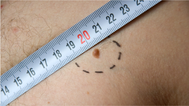
- The cancer is more than 2 mm thick in diameter
- It has grown into the lower dermis
- It has grown into the space around a nerve
- It started to spread to the ear or lip
- The cells are poorly differentiated or undifferentiated when seen in a microscope
Clark and Breslow staging:
The stage of melanoma is also determined by how deeply the cancer has grown into the skin and if it has spread and there are two scales commonly used to describe how deep the melanoma has gone into the skin.
Clark scale:
The Clark scale has five levels:
Level 1: The melanoma cells are only in the outer layer of the skin, the epidermis
Level 2: The cancer cells are directly beneath the epidermis in a layer called papillary dermis
Level 3: The cancer cells have spread through the papillary dermis to reticular dermis
Level 4: The melanoma cells have spread through the reticular or deep dermis
Level 5: The cancer has grown into the layer of subcutaneous fat
Breslow scale:
In the Breslow scale, the doctor measures the thickness of the melanoma using a micrometer and this is called the primary tumour thickness or Breslow thickness.
Stages of non melanomas:
Stage 0:
Also called carcinoma in situ or Bowen’s disease, this stage can be considered precancerous. The cells affected by cancer are in the same location (in situ) and have not spread to through the tissue.
Stage I:
The cancer is 2 cm across or lesser and has one or no high risk features.
Stage II:
The cancer is 2 cm across and has two or more high risk features.
Stage III:
The cancer has grown into into the bones in the face, like the jaw bone or the bone around the eye or it has spread to a nearby lymph node than it less than 3 cm in size.
Stage IV:
The cancer has spread into the spine, lower part of the skull or the ribs or it has spread to a lymph node more than 3 cm in size or to an internal organ such as the lungs.
Grade:
The grade describes how closely the cancer cells resemble the normal cells of the skin when seen under a microscope. The scaling is from 1 to 3 in grading skin cancer:
Grade 1 (G1): The cancer cells look like the normal skin cells
Grade 2 (G2): The cells look somewhat in between grade 1 and grade 3
Grade 3 (G3): The cancer cells look abnormal and unlike the normal cells