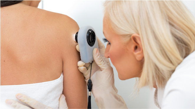
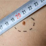
The symptom of skin cancer is very often a skin lesion, except for advanced stages. Most patients who examine their own skin regularly may spot suspicious skin changes and report them. This is what happens in at least 40% of the cases. Dermatologists examine the changed skin parts using a dermatoscope and evaluate the risk factors of the patient acquiring a skin cancer and proceed to diagnosis. Read more about skin cancer here.
Biopsy:
A common test to diagnose a skin cancer is a skin biopsy. A skin tissue is extracted and sent to a laboratory for pathological study. A biopsy not only identifies a cancer’s skin tissue but also helps in diagnosing the stage of cancer. A local anesthesia is administered while performing skin biopsies. There are many types of biopsies available, the exact type to be performed over a particular patient depends upon the the size of skin, location among other factors. Following are the types of biopsies used for confirming skin cancers:
Shave (tangential) biopsy:
The top layers of the skin are shaved off using a surgical blade for a biopsy. This type of biopsy is usually used to diagnose may skin diseases. For a skin cancer diagnosis, this not used when a melanoma is suspected very strongly, as the blade may not be of enough thickness to measure the depth of cancer invasion.
Punch biopsy:
A tiny cutter like tool is used in punch biopsy to extract a sample of deeper skin tissues. The tool is rotated through all layers of skin and the ends of the cuts are often stitched up.
Incisional or excisional biopsies:
In a incisional or excisional biopsy a knife is used to reach the entire thickness of the skin and sample is extracted. It is used when the cancer have grown into deep layers of skin, the ends of skin are stiched up for healing after biopsy. Excisional biopsy removes the whole tumor as such and so is regularly practiced and preferred method for strong suspicions. The incisional biopsy only removes a part of the tumor.
Optical biopsies:
Optical biopsy is relatively new type of biopsy using machines like reflectance confocal microscopy (RCM). This doesn’t require the removal of skin samples.
Biopsies for melanoma that might have spread:
Skin cancer can also spread to other organs of the body like lungs and lymph nodes. This can happen after the appearance of a skin lesion or while it is developing or even after its disappearance. These kind of tumors are found using imaging test such as CT scan or a different kind of biopsies are performed.
Fine needle aspiration (FNA) biopsy:
A thin syringe needle is used to remove pieces of lymph nodes and this is only used to find out if the melanoma has spread to nearby lymph nodes but not on suspicious moles. A CT scan is used to guide the needle to the site of tumor. The amount of sample collected through this method may not be sufficient to gather all information about the tumor though.
Surgical (excisional) lymph node biopsy:
A large portion of the lymph node is removed surgically through a cut using anesthesia in this method of biopsy. This is often used as a follow up to FNA after finding that the cancer has spread.
Sentinel lymph node biopsy:
This is used after knowing specific details about the cancer to see if the cancer has spread to the lymph nodes nearby and if it will affect the treatment option chosen. A radioactive substance is injected in the site of the melanoma tumour. The radioactive substance collects in one or more sentinel nodes viewed by a small camera. Once these are marked by injected dye in the same area as the radioactive substance a small incision is performed and the nodes are removed. If melanoma cells are not found a surgical procedure is not necessary. If a sentinel node is swollen or enlarged and hard with mid tumor thickness greater than 1 millimeter in size a sentinel node biopsy may not be required, a normal biopsy would suffice.
Imaging Tests:
Imaging tests are often used to see the possible metastasized melanoma cells in the body using X rays, radioactive substances, magnetic fields to create pictures of the body from inside.
This is done in advanced stages of cancer only. It helps to determine the effectiveness of treatment and find recurring cancers. These tests include:
Chest x-ray:
If melanoma has spread to lungs it is determined using the chest X ray test.
Computed tomography (CT) scan:
CT scan specializes in determining the changes in the soft issues, that is, in organs of the body. A complete cross sectional image of lymph nodes is examined to see any enlargement or suspicious spots of lymph nodes.
Magnetic resonance imaging (MRI) scan:
Radiowaves and strong magnets are used in an MRI scan to get detailed images especially of brain spinal cord.
Positron emission tomography (PET) scan:
A radioactive form of sugar is injected to collect cancer cells. A camera is employed to take pictures of areas of radioactivity in the body. This is usually used only in advanced stages of skin cancer.
PET-CT scan:
A combination of PET and CT scan is done usually so that the potential cancerous areas are found out and their intensity is checked at the same time.
Blood tests:
Blood test may not directly diagnose the presence of a cancer, however the extent of spread can be diagnosed by looking at the blood picture of a patient. The working condition of bone marrow, liver, and kidneys is determined using the blood chemistry levels in patients with advanced stages of melanoma.The level of LDH(Lactate Dehydrogenase) is determined to check if melanoma has spread to distant parts of the body and to determine the stage of cancer A high LDH means that the cancer is difficult to treat.


