
Retinoblastoma is a type of childhood cancer that starts in the back portion of the eye known as retina. In children, this cancer is the most common of all eye cancers.
 Each year, close to 20 percent of the global retinoblastoma cases are diagnosed in India. Of these, between 10 and 30 percent are able to lead functioning lives into adulthood. While a majority of retinoblastoma cases are found in the rural regions, many are unaware of the condition and are diagnosed usually in advanced stages or, despite early diagnosis are not able to afford treatment.
Awareness and early detection are key to increase cure rates and survival rates.
The eye and retina
 The eyeball is the eye’s main part and is filled with vitreous humour – a jelly-like substance. The eyeball, on its front, has a clear lens with iris which is the coloured portion of the pupil. The retina is located in the back part of the eye and consists of a layer of nerve cells that are sensitive to light. The optic nerve goes out of the back of the eyeball and connects the retina to the brain.
Light enters the eye through the lens and is then focused on the retina. The image or pattern of light that is focused on the retina is sent by the optic nerve to the visual cortex – a special region in the brain that allows us to see.
How does retinoblastoma occur?
The eyes develop much before birth. In the early stages, the eyes have retinoblasts that are cells which multiply and form new cells that make up the retina. At some point, these cells become mature cells and stop multiplying.
At times, this process goes wrong and instead of maturing, retinoblasts grow rapidly out of control to form a tumour called retinoblastoma.
Although a complex chain of events occurs to cause retinoblastoma, a mutation in a gene known as RB1 gene is commonly associated with this type of cancer. While the RB1 gene normally prevents uncontrolled growth of cells, mutation in the gene hampers its functioning. Based on where and when the RBI gene mutation happens, retinoblastoma is classified into two different types.
- Congenital retinoblastoma
- Sporadic retinoblastoma
Congenital retinoblastoma
The RBI gene mutation is present at birth (congenital) in one out of three children with retinoblastoma. This genetic abnormality is present in all the body cells including cells in the retinas of both eyes. This type of mutation is also called germline mutation.
Although called hereditary or ‘heritable’ cancer, no family history is seen in many children and RB1 gene mutation is not inherited from parents. In these children, the mutation occurs first during the early development stages in the womb. Very few children inherit the gene mutation from one of the parents.
Children who have RB1 gene mutation develop retinoblastoma usually in both eyes where it is known as bilateral retinoblastoma and several tumours are seen within the eye (multifocal retinoblastoma).
As the RBI gene mutation is present in all body cells, these children are at higher risk for other cancers as well.
Some children with congenital (present at birth) retinoblastoma may develop another tumour in the base of the brain, typically in the pineal gland and this type of cancer is called pineoblastoma or trilateral retinoblastoma.
Children who are survivors of hereditary retinoblastoma have a higher-than-average risk of developing other cancers in adulthood.

Sporadic retinoblastoma
In two out of three children, RB1 gene abnormality develops in one cell only in one eye.
Only one eye is affected with the tumour in a child with sporadic retinoblastoma. Sporadic retinoblastoma is usually found in slightly older children as compared to the heritable form.
The risk of developing other cancers is lower in children with sporadic retinoblastoma.
Symptoms of retinoblastoma
As retinoblastoma affects infants and children, symptoms are rare and not noticed. Signs that may be noticed can include
- Redness in the eye
- Swelling of the eye
- Poor vision
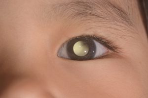
- White colour in the pupil or centre of the eye when exposed to light such as during a flash photograph
- Enlarged pupil
- Infection in the eye
- Eyeball looking larger than normal
- Cloudy pupil and iris (the coloured part of the eye)
Growth and spread of retinoblastoma
If untreated, retinoblastomas grow and fill most of the eyeballs. From the main retinal tumour, cells can break away to reach other regions of the eye to form more tumours. These tumours can affect the normal fluid circulation in the eye, raising the eye pressure. This causes a condition called glaucoma, leading to loss of vision and pain.
Most retinoblastomas, however, are treated before they metastasize out of the eyeball. If they are not treated retinoblastoma spreads to other parts and can reach the brain by growing along the optic nerve. Retinoblastoma cells may grow into eyelids, eye socket and nearby tissue and affect nearby lymph nodes or distant organs including liver, bone marrow or bones.
Diagnosis of retinoblastoma
Retinoblastoma is diagnosed with the following tests or procedures:
Eye exam
The eye doctor performs an eye exam to know the cause behind the signs and symptoms.
Imaging tests
Imaging tests and scans help determine whether retinoblastoma has affected other structures of the eye. Imaging tests include computerized tomography (CT), ultrasound, and magnetic resonance imaging (MRI).
Some other diagnostic tests that may be used include:
Ophthalmoscopy
involves checking the optic nerve and the retina with a small magnifying lens.
Slit-lamp biomicroscopy
A microscope and strong light are used to examine optic nerve, retina and other eye parts.
Fluorescein angiography
This is a procedure where fluorescein, an orange fluorescent dye is used to check the flow of blood and blood vessels inside the eye.
What are the treatment options for retinoblastoma?
Treatment options depend on the location and size of the tumor and whether other areas are affected and the child’s overall health.
Chemotherapy
Chemotherapy uses drugs to kill cancer cells and helps shrink a tumour in order to improve the effectiveness of other treatments such as radiation, laser therapy or cryotherapy. These treatments when used in combination may help avoid surgery.
Chemotherapy is also the treatment option when the cancer has spread to other body parts or tissues that lie outside the eyeball.
Intra-arterial chemotherapy is a new type of treatment that directly delivers the drug to the tumour using a tiny tube in the eye. This chemotherapy may be used as an initial treatment for retinoblastoma that does not respond to other treatments.
Radiation therapy
Radiation therapy uses X-rays, to destroy cancer cells.
Internal radiation or brachytherapy for retinoblastoma involves the use of a small disk that has radioactive material. The disk is directly stitched in the affected place where it remains for a few days slowly giving off radiation to cancer cells.
External beam radiation involves directing high-powered beams from a machine placed externally to the tumour.
Laser therapy
Laser therapy or laser photocoagulation uses laser beams to destroy blood vessels which provide nourishment in the form of nutrients and oxygen to the tumour. Cancer cells die when there is no nourishment.
Cryotherapy
Cryotherapy involves using extreme cold to destroy cancer cells.
Liquid nitrogen or other cold substances are placed near or directly into the cancer cells. The extreme cold enables cancer cells to freeze after which liquid nitrogen is removed, allowing the cells to thaw. Freezing and thawing is repeated in each session of cryotherapy to cause death of cancer cells.
Surgery
Surgery is the treatment option when the cancer is too large to respond to other methods. Surgery is used to remove the affected eye to help prevent the cancer from spreading to other body parts.
Surgery involves:
Enucleation or removal of the affected eye – In this surgery, the muscles and tissue surrounding the eye are disconnected and the eyeball is removed. A part of the optic nerve that connects the back of the retina to the brain is also removed.
Eye implant surgery – After removing the eyeball, an implant that is made of plastic or some other material is placed by the surgeon in the socket of the eye and the eye muscles are connected to the implant. Although the child cannot see with the implant, the eye muscles move normally after they adapt to the implant.
Artificial eye – Several weeks post surgery, an artificial eye that is custom-made to match the healthy eye may be placed on the eye implant.
Genetic counselling
Genetic counselling is an important aspect of the treatment of the two types of retinoblastoma. Genetic counselling involves a discussion that parents have with a trained healthcare professional to understand genetic diseases and to test for RBI gene mutation. Retinoblastoma risk for the child as well as brothers or sisters is also discussed in genetic counselling sessions.
Children who have a known retinoblastoma family history should have regular eye exams that begin early in life to check for the condition.
Siblings of a child diagnosed with retinoblastoma should also undergo regular eye exams by the ophthalmologist until they reach the age between 3 to 5 years.
If you or your loved one has any symptoms listed above or would like to know more about retinoblastoma, the first step is to consult a doctor. Book an appointment with a top oncologist in your area at www.onco.com.