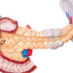
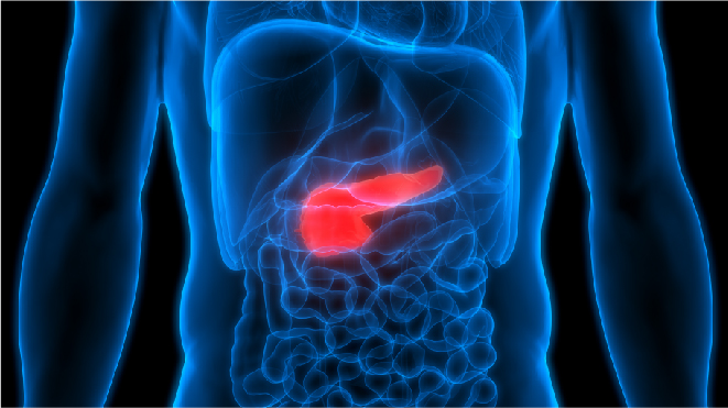
Gallbladder cancer as the name suggests, begins in the gallbladder, an organ that stores a substance called bile. To read about gallbladder and gallbladder cancer, click here
How is gallbladder cancer diagnosed?
Sometimes gallbladder cancer is detected due to gallstones or inflammation of the gallbladder or during surgical procedures for the same though most cases are found out until the patient goes to the doctor with the presented symptoms, and the following tests may be used to diagnose gallbladder cancer.
Medical history and physical exam:
If the doctor suspects the patient to have gallbladder cancer, a complete medical history is taken to check for risk factors and symptoms that can point out to the cancer. A physical examination may include checking the abdominal area for lumps, tenderness or fluid buildup. Eyes and skin may be checked to see if there are any signs of jaundice.
Liver function tests:
Lab tests may be done to find out the levels of bilirubin, albumin, liver enzymes such as alkaline phosphatase, AST, ALT and GGT and certain other substances in the blood and these are collectively called liver function tests and they can help diagnose gallbladder cancer and related conditions, liver or bile duct disease.
Tumour marker tests:
A blood sample is taken and is tested for biomarkers that can be elevated in the presence of a particular type of cancer and this procedure is called tumour marker test. People with gallbladder cancer may have high levels of markers called CEA and CA 19-9, though these are not unique to gallbladder cancer and are high only in the advanced stages. Prior to any treatment, the levels of the serum tumour marker can be taken and compared to the tumour marker levels after to see the patient’s response to treatment.
Ultrasound:
This procedure uses ultrasounds which bounce back on internal tissues or organs in the neck, and make echos. These echoes give a picture of the organs,also known as sonograms. Ultrasounds are used during biopsies too, to guide the fine-needle aspiration.
MRI:
Magnetic resonance imaging uses radio waves and strong magnets for imaging and it provides great detail and is useful to image gallbladder, nearby bile ducts and other organs.
CAT or CT scan:
Also known as tomography, computerized tomography, or computerized axial tomography, this scan gives the detailed pictures of the organs from different angles, generated by a computer which is linked to an x-ray machine. For the scan, a dye is given to the patients intravenously or orally for the tissues to be imaged clearly.
Cholangiography:
This is an imaging test to check if the bile ducts are blocked, narrowed or dilated and it is used to plan for surgeries to treat gallbladder cancer. Magnetic resonance cholangiopancreatography (MRCP), endoscopic retrograde cholangiopancreatography (ERCP) and percutaneous transhepatic cholangiography (PTC) are the types of cholangiograms available to diagnose gallbladder cancer.
Angiography:
Angiogram is an X ray used to look at the blood vessels of the patient. A thin plastic tube is inserted into the artery and a small amount of contrast dye is injected to outline the blood vessels. This can show the tumours or blockages in the blood vessels that can be noted if the cancer has spread beyond the gallbladder into the surrounding blood vessels.
Laparoscopy:
Laparoscope is a thin tube with a light and a video camera than is inserted into the patient’s abdomen through a small incision, to look at the gallbladder, the liver and nearby organs and if needed a biopsy can be done too to confirm cancer. Laparoscopy can help determine the stage and extent of gallbladder cancer too.
Biopsy:
Biopsy for involves the removal of cells from the suspicious areas and examining them under a microscope and is not the prefered method of diagnosis for gallbladder cancer. This is because in case of gallbladder cancer, it can cause the cancer to spread and even if required, surgical biopsy is the prefered option, it means examination of cells removed through or post surgery.
How is gallbladder cancer staged?
Staging of gallbladder cancer helps the doctors figure out how much the cancer has spread in the pancreas and determine its best treatment. Staging also helps calculate survival statistics. The lower the number of the stage, the less is the cancer has spread, with early stages being 0 called carcinoma in situ, followed by stage 1 and the most advanced stage being stage 4. The following are the factors taken into consideration for staging of gallbladder cancer:
TNM method:
Cancer types that form tumours are staged using TNM system and the same method is used for gallbladder cancer too. The extent of the primary tumour (T), the presence of cancerous lymph nodes (N) and how far the gallbladder cancer has spread to a different part of the body (M) can be described using the TNM system.
Grade:
The grade describes how closely the cancer cells resemble the normal cells of the gallbladder when seen under a microscope. The scaling is from 1 to 3 in grading gallbladder cancer:
Grade 1 (G1): The cancer cells look like the normal gallbladder cells
Grade 2 (G2): The cells look somewhat in between grade 1 and grade 3
Grade 3 (G3): The cancer cells look abnormal and unlike the normal cells
Extent of resection:
Resection means removal of the cancer tumour through surgery, and based on the extent of resection the gallbladder cancers can be classified as:
Resectable: Those that the doctors believe can be completely removed by surgery
Unresectable: Those that have spread too far or in a location that cannot be operated on through surgery.
Only a small number of gallbladder cancer cases are resectable even when first diagnosed. The exact staging of gallbladder cancer and characteristics of each stage are discussed in the further articles.
Stage I of gallbladder cancer:
Stage II of gallbladder cancer:
Stage III of gallbladder cancer:
Stage IV of gallbladder cancer: