What are the screening systems for bone cancer?
Imaging tests and blood tests might suggest that a person has bone cancer. In most cases, however, doctors confirm this through a biopsy, which is a procedure that tests a tissue or cell sample by checking it under a microscope.
Imaging Tests:
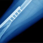
1. X-ray:
A diagnostic test that uses invisible electromagnetic energy beams to make images of internal tissues, bones, and organs onto film. The bone at the affected area may look disorganized and ragged instead of solid. Cancer may also appear as a hole in the bone. It is also possible that the tumor spread to nearing muscle, fat, or organs.
The radiologist will mostly be able to tell the severity of the condition by looking at the X-ray images, but a biopsy is mandatory to confirm.
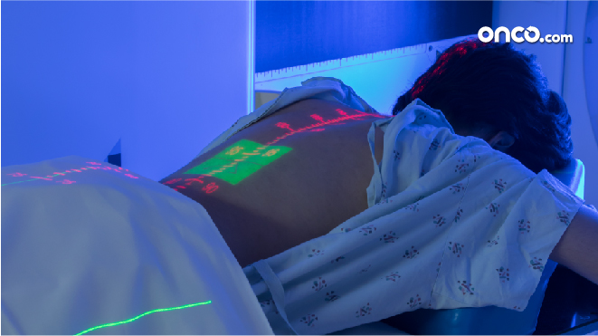
Computed tomography scan (also called a CT or CAT scan):
It is an imaging test that uses X-rays and a computer to make detailed images of the body. A CT scan shows details of the bones, muscles, fat, and organs. It is helpful in staging cancer.
It is used to guide a biopsy needle into a tumor which is known as a needle biopsy. For this, the individual is inspected on the CT scanning table while the radiologist moves the biopsy needle towards the tumor, and CT scans shots repeat until the needle is inside the tumor.
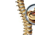
2. Magnetic resonance imaging (MRI):
A diagnostic procedure that uses a combination of large magnets, radio frequencies, and a computer to make detailed images of organs and structures within the body. It is a powerful magnet link to a computer to take detailed pictures of the tumor without using X-rays. Also, it is most useful in gauging tumors near the brain or the spinal cord.
3. Positron emission tomography (PET) scan:
An imaging test in which radioactive-tagged glucose (sugar) enters the bloodstream. Tissues that a scanning machine can consume glucose more than healthy tissues (such as tumors). As cancer cells use more glucose than healthy cells, the images spot cancer cells in the body.
4. Biopsy:
In a biopsy, tissue samples are removed with a needle or during surgery from the body for examination under a microscope. It is done to determine if cancer or other abnormal cells are present. An experienced surgeon does the biopsy.
a. Needle Biopsy
A needle biopsy is of two types (fine aspiration), and core, both of which happen after the affected area is numbed.
b. Surgical Biopsy
There are two types of surgical biopsy, one, incisional biopsy and two, excisional biopsy. While the former cuts through the skin to more a small piece of tissue, the latter removes the entire tumor.