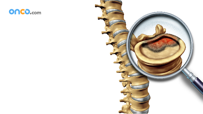
How is the diagnosis of bone cancer made?
Accurate diagnosis of bone cancer include the combination of information on the bone affected, then part of the bone involved, X-rays, and a biopsy. While other diseases like bone infections can cause symptoms and imaging results for them can cause misdiagnosis.
Imaging tests used for diagnosis:
1. X-ray:
A diagnostic test that uses invisible electromagnetic energy beams( X-ray) to make images of internal tissues, bones, and organs onto film. The bone at the affected area may look disorganized and ragged instead of solid. It is also possible that the tumor spread to nearing muscle, fat, or organs.
An experienced radiologist may be able to diagnose the condition by looking at the X-ray images, but a biopsy is mandatory to confirm.
2. Computed tomography scan (also called a CT or CAT scan):
It is an imaging test that uses X-rays and a computer to make detailed images of the body. A CT scan shows details of the bones, muscles, fat, and organs.
It is also used to guide a biopsy needle into a tumor which is known as a needle biopsy. For this, the individual is inspected on the CT scanning table while the radiologist moves the biopsy needle towards the tumor, and CT scans shots repeat until the needle is inside the tumor.
3. Magnetic resonance imaging (MRI):
A diagnostic procedure that uses a combination of large magnets, radio frequencies, and a computer to make detailed images of organs and structures within the body. It is a powerful magnet link to a computer to take detailed pictures of the tumor without using X-rays. It is most useful in gauging tumors near the brain or the spinal cord.
4. Positron emission tomography (PET) scan:
An imaging test in which radioactive-tagged glucose (sugar) enters the bloodstream. A scanning machine can detect tissues that use glucose more than healthy tissues (such as tumors). As cancer cells use more glucose than healthy cells, the images spot cancer cells in the body.
5. Biopsy:
A procedure in which tissue samples are removed with a needle or during surgery from the body for examination under a microscope. It is done to determine if cancer or other abnormal cells are present. Also, an experienced surgeon/physician must do a biopsy.
a. Needle Biopsy
A needle biopsy is of two types (fine aspiration), and core, both of which happen after the affected area is numbed.
In fine needle aspiration (FNA), the surgeon uses a thin needle and a syringe to extract some fluid and some cancer cells. If the tumor lies close to the surface of the individual’s skin, the surgeon may aim the needle by feeling the region. However, if it is deep, the doctor may guide the needle by looking at the CT scan pictures. An X-ray specialist or an interventional radiologist conducts a CT guided biopsy.
In a core needle biopsy, the doctor uses a larger needle which is about 1/16 inches in diameter and ½ inches long to remove a small cylinder of tissue. Experts recommend core needle biopsy over fine needle biopsy to diagnose primary bone cancer.
b. Surgical Biopsy
There are two types of surgical biopsy. One, an incisional biopsy and two, an excisional biopsy. While the former cuts through the skin to more a small piece of tissue, the latter removes the entire tumor.